
Fig. 4.
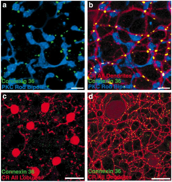
Cx36 immunoreactivity in retinal whole-mounts. a: Rod bipolar cells, although sometimes stained diffusely by the Cx36 antibody in the somatic and axonal regions, are not stained at all at their terminals, which are stained by an antibody to protein kinase C (PKC, blue). Some Cx36-immunoreactive puncta (green) do appear nearby, but rarely appear on the terminals. b: The dendrites of AII amacrine cells, stained by anti-calretinin (CR, red), are intimately associated with the Cx36 puncta. The few colocalizations of rod bipolar cell terminals with Cx36 (e.g., lower right) can invariably be accounted for by passing AII amacrine cell processes. c: Cx36 immunoreactivity is sparse and faint in sublamina a. The puncta that are found do not colocalize with the lobules of AII amacrine cells, which do not make gap junctions in this area. d: In the same field as c, focus on sublamina b demonstrates the extent to which Cx36-immunoreactive puncta are located on AII amacrine cell dendrites. Scale bars = 10 μm in a,b, 20 μm in c,d.