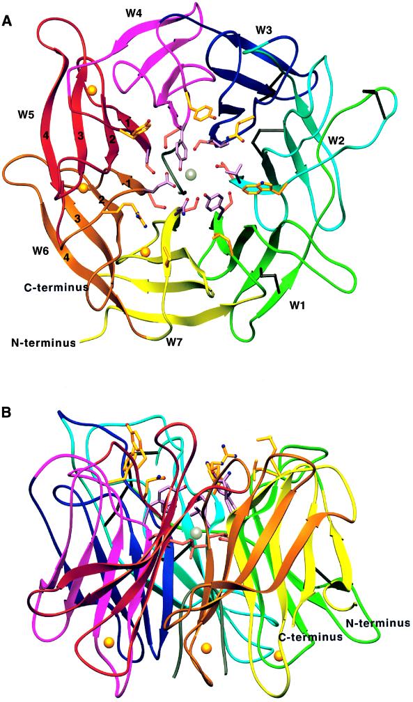
Figure 3.
Ribbon diagrams (65) of the model for the integrin α4-subunit β-propeller domain. Views are from the top (A) and side (B). Each W is shown in a different color. A hypothetical polypeptide finger in the central cavity is gray. Cysteines in disulfides are black. Side chains in β-strand 1 in positions 0, b, and 2 are shown in gold, lavender, and rose, respectively; their oxygens and nitrogens are red and blue, respectively. Ca2+ ions and a hypothetical Mg2+ ion are gold and silver spheres, respectively.

