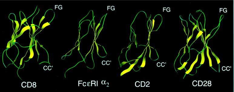
Figure 6.
Surface-binding pockets consisting of FG and CC′ loops observed in: CD8α, which mediates dimerization (35); FcɛRIα2, which is the domain of IgE high- affinity receptor that binds IgE (36); CD2, which binds LFA-3 (37); and CD28, which binds CD80/86 (38). These surface interaction sites show a similar feature to that of the CD4 protein as shown in Fig. 1. The structure of FcɛRIα2 was modeled based on the crystal structures of CD2 domain 2 (37) and CD4 domain 2 (12). The structure of CD28 was modeled by using the crystal structure of the VH chain of HYHEL-5 Fab (39).

