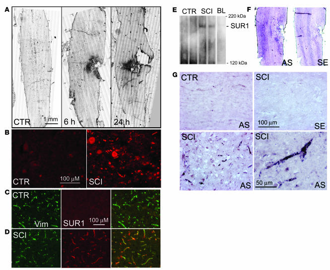
Figure 1. SUR1 is upregulated in SCI.
(A) Immunohistochemical localization of SUR1 in control rats (CTR) and at different times after SCI as indicated, with montages constructed from multiple individual images and positive labeling shown in black pseudocolor. (B) Magnified views of SUR1-immunolabeled sections taken from control and from the core (heavily labeled area in A at 6 hours). (C and D) Immunolabeling of capillaries with vimentin (Vim) and colabeling with SUR1 in control rats (C) and from the penumbra of SCI rats (tissue adjacent to the heavily labeled core in A, 6 hours) (D). (E) Western blots for SUR1 of spinal cord tissue from control rats (50 μg protein), from rats 6 hours after SCI (50 μg protein), and from an equivalent amount of blood (BL; 2 μl) as is present in the injured cord. Blots are representative of 5–6 control and SCI rats. (F and G) In situ hybridization for Abcc8 in control rats and in whole cords (F) or in the penumbra (G) 6 hours after SCI using antisense (AS) and sense (SE) as indicated. Immunohistochemistry and in situ hybridization images are representative of findings in 3–5 rats per group. Scale bars: 1 mm (A); 100 μM (B–D and G, top panels and bottom left panel); 50 μM (G, bottom right panel).

