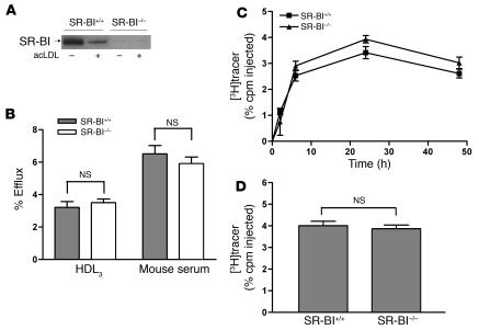
Figure 2. SR-BI–deficient BMMs have normal cholesterol efflux ex vivo and RCT in vivo.
(A) Western blotting demonstrating the absence of SR-BI protein expression in SR-BI–/– macrophages. SR-BI was detected by Western blotting with anti–SR-BI antibody. Equal amounts of total proteins were loaded. (B) Mouse BMMs from WT (SR-BI+/+) and SR-BI–/– mice were labeled with [3H]cholesterol for 24 hours. After equilibration in 0.2% BSA overnight, macrophages were incubated with either 25 μg/ml HDL3 or 2.5% mouse whole serum for 2 hours. Values are mean ± SD; n = 3. Results are representative of 2 independent experiments. (C and D) The RCT experiment was performed as described in Figure 1 for SR-BI+/+ and SR-BI–/– BMMs, except that cells were not loaded with acLDL and neither cells nor mice were treated with GW3965. n = 6 mice per group. Data are expressed as the percentage of tracer relative to total cpm tracer injected ± SEM. Results are representative of 2 independent experiments. (C) Time course of [3H]cholesterol distribution in plasma. (D) Fecal [3H]tracer levels. Feces were collected continuously from 0 to 48 hours.

