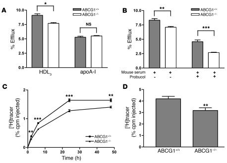
Figure 5. ABCG1-deficient BMMs have reduced cholesterol efflux ex vivo and RCT in vivo.
(A) Cholesterol efflux assay was determined as described in Figure 1 with BMMs from WT (ABCG1+/+) or ABCG1-KO (ABCG1–/–) mice. Cholesterol efflux was determined in the presence of HDL3 (25 μg/ml) or lipid-free apoA-I (10 μg/ml) for 2 hours. Data are expressed as mean ± SD; n = 3. *P < 0.05. (B) Cholesterol efflux was determined as described above, except cells were incubated for 2 hours either in the presence or absence probucol (20 μM) prior to the addition of 2.5% mouse whole serum as the acceptor. Data are expressed as mean ± SD; n = 3. **P < 0.01; ***P < 0.001. (C and D) The RCT assay was performed as described in Figure 1 with [3H]cholesterol-labeled, acLDL-loaded, and LXR agonist–treated BMMs from ABCG1+/+ or ABCG1–/– mice. n = 8 mice per group. Data are expressed as the percentage of tracer relative to total cpm tracer injected ± SEM. **P < 0.01; ***P < 0.001. Results are representative of 2 independent experiments. (C) Time course of [3H]cholesterol distribution in plasma. Individual time points and areas under the curve were determined and compared. (D) Fecal [3H]tracer levels. Feces were collected continuously from 0 to 48 hours.

