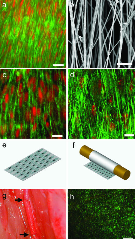
Fig. 1.
Characterization of biomimetic cell–scaffold interactions. (a) En face staining of SMCs in a native rat CCA after EC denudation. The samples were stained for actin filaments by using FITC-conjugated phalloidin (green), and they were counterstained for nuclei by using propidium iodide (red). (Scale bar, 20 μm.) (b) SEM of aligned nanofibers made from PLLA. (Scale bar, 10 μm.) (c) Human aortic SMCs seeded on aligned nanofiber surface, with same staining as in a. (Scale bar, 20 μm.) (d) MSCs seeded on aligned nanofiber surface, with same staining as in a. (Scale bar, 20 μm.) (e) Schematic illustration of cells seeded on a piece of nanofibrous membrane coated with fibronectin. (f) Embedding cells in the tubular grafts by rolling the nanofibrous membrane. (g) An end-to-end anastomosis procedure of the vascular graft sutured to the CCA in a rat. The arrows indicate the two ends of the graft. (h) Cell survival in a vascular graft after surgery process. En face live/dead staining of a TEVG was performed to show calcein-positive (live) cells in green and ethidium bromide-positive (dead) cells in red. (Scale bar, 200 μm.)

