
Figure 5.
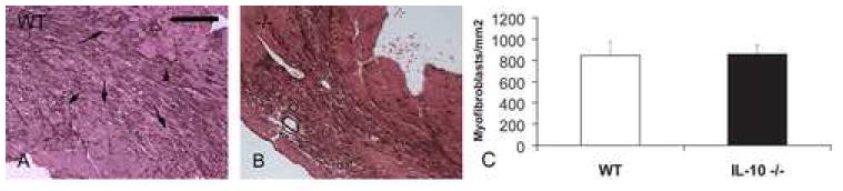
Myofibroblasts are identified in the healing infarct as extravascular α-SMA-expressing spindle-shaped cells. A. After 72 h of reperfusion dead cardiomyocytes are replaced with granulation tissue, and myofibroblasts infiltrate the infarcted area, predominantly localized in the border zone (arrows). B. IL-10 null mice show comparable myofibroblast accumulation in the infarct. C. Quantitative analysis of myofibroblast density in the infarcted myocardium (scale bar=70 μm).