Figure 4.
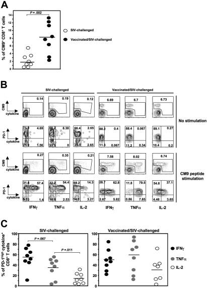
PD-1highCM9+CD8+ T cells are capable of producing multiple cytokines. (A) Dot-plot graph showing the percentage of CM9+CD8+ T cells in PBMCs from SIV-challenged (n = 8) and vaccinated/SIV-challenged (n = 8) animals. (B) Representative flow cytometry showing the production of IFNγ, TNFα, and IL-2 by CM9+CD8+ T cells after 6-hour stimulation with CM9 peptide. Single-live lymphocytes were gated first for CD8- and then for CM9-specific CD8+ T cells by CM9 tetramer. The production of all 3 cytokines was simultaneously measured in CM9+CD8+ T cells and in relation to their PD-1 expression. Cells from one SIV-challenged (left panel) and one vaccinated/SIV-challenged (right panel) animal for both no stimulation and CM9 stimulation conditions are shown. (C) Compiled data showing the percentage of PD-1highCM9+CD8+ T cells that is positive for each one cytokine tested in SIV-challenged (left panel, n = 8) and vaccinated/SIV-challenged (right panel, n = 8) animals. Horizontal lines depict mean values. The P values were calculated using Student t test.

