Figure 3.
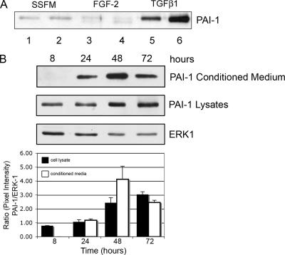
PAI-1 is secreted in response to TGFβ1. (A) Western blot for PAI-1. Fibroblasts were plated in SSFM, SSFM-FGF, or SSFM-TGFβ. At 72 h, conditioned media were collected and subjected to Western blot detection for PAI-1. Duplicates represent media from cells cultured from two different cornea donors. PAI-1 is labeled. TGFβ1 induced PAI-1 secretion, whereas FGF-2 did not. (B) Cells were plated in SSFM-TGFβ and grown for 8, 24, 48, or 72 h. At the specified time points, cells were lysed. Both conditioned media and lysates were detected for PAI-1 secretion and expression by Western blot. In the cells treated with TGFβ1, PAI-1 was detected in cell lysates at 8 h and in conditioned media at 24 h; expression increased with time. ERK1 expression functions as a control for protein loading. The ratio of the pixel densities (PAI-1/ERK1) is calculated and graphed. The Western blot image is representative of three independent experiments.

