Figure 6.
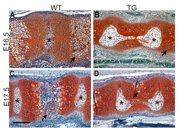
Safranin O stainings for cartilage of mid-sagittal sections through the presumptive vertebrae of transgenic and wild-type embryos. (A) In wild-type embryos at E16.5 hypertrophic chondrocytes are observed in the centre of the vertebrae (arrow). (B) The number of chondrocytes undergoing hypertrophic differentiation is reduced in transgenic embryos (arrow). At E17.5 (C) chondrocytes of wild-type embryos lose the cartilage matrix and vascularisation and invasion of osteocytes starts (arrow). The notochord is extruded from the vertebrae. (D) In transgenic vertebrae of the same age fewer cells are hypertrophic and are dorsally displaced (arrow). Ossification is delayed and notochord extrusion is incomplete. Scale bar: 200 μm. Asterisks: nucleus pulposus.

