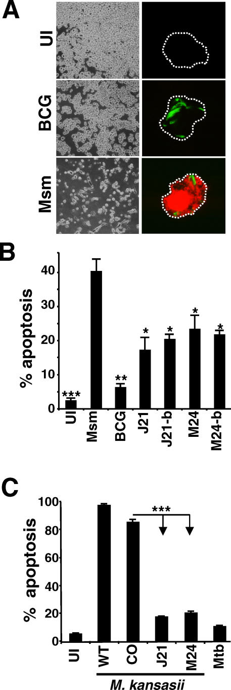
Figure 1. Identification of Regions in the Mtb Genome Mediating Inhibition of THP-1 Cell Apoptosis.
(A) M. smegmatis (Msm) induced more cell death in infected THP-1 cells than BCG or uninfected cells (UI) as observed by bright field microscopy (left panels) and fluorescence microscopy of TUNEL staining (right panels; red fluorescence is TUNEL staining, and green fluorescence is GFP-labeled bacteria).
(B and C) M. smegmatis was transfected with an episomal cosmid library of Mtb genomic DNA, and individual clones were screened for their capacity to inhibit apoptosis. The cosmid DNA of two selected clones (J21 and M24) was purified and used to re-transfect M. smegmatis, resulting in clones J21-b and M24-b. These clones, along with the original transformants (M24 and J21), uninfected (UI) cells, M. smegmatis (Msm), and BCG were tested for induction of apoptosis by infection of THP-1 cells followed by TUNEL staining at 16 h after infection and flow cytometry (C). The J21 and M24 cosmids and empty vector cosmid (CO) were transfected into M. kansasii, and the induction of apoptosis by the bacteria was compared to uninfected and Mtb-infected THP-1 cells using a TUNEL assay as in (B), except that cells were harvested after 5 d of infection. Results in (B and C) are averages of three independent experiments and error bars represent ± standard deviation (SD). Statistical significance relative to levels of apoptosis induced by wild-type M. smegmatis in (B) or M. kansasii in (C) is indicated as follows: *, 0.01 < p < 0.05; **, 0.001 < p < 0.01; ***, p < 0.001 (ANOVA with Tukey post-test).

