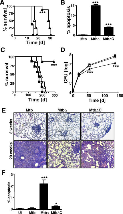
Figure 4. Identification of nuoG as a Mycobacterial Virulence Determinant.
(A) Survival of SCID mice after intravenous infection with 106 wild-type Mtb (squares), ΔnuoG mutant (filled triangles), or complemented mutant bacilli (open triangles). n = 7 mice per group.
(B) Apoptotic cells in lung tissues of SCID mice after 14 d of infection were quantified using TUNEL peroxidase staining by microscopy as explained in Figure 2.
(C) Survival of immunocompetent BALB/c mice infected with bacterial strains as in (A); n = 7 mice per group.
(D) The bacterial burden in the lungs of infected BALB/c mice was followed (n = 3 per time point; symbols indicate bacterial strains as in [A and C]).
(E) Lung histopathology (hematoxylin and eosin staining) at 3 and 20 wk for BALB/c mice infected as in (C) with wild-type Mtb, ΔnuoG mutant (MtbΔ), or complemented mutant Mtb (MtbΔC).
(F) The percentages of apoptotic cells in lung sections were determined by TUNEL peroxidase assay and quantified in blinded fashion as in Figure 2. All results shown are representative of two independent experiments. Statistically significant differences compared to wild-type Mtb are indicated by asterisks as in the Figure 1 legend.

