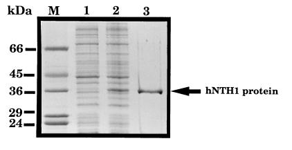
Figure 3.
Purification of recombinant hNTH1 protein. A Coomassie blue-stained SDS/polyacrylamide gel is shown of a lysate of BL21 cells containing the pET14b/hNTH1 plasmid before (lane 1) and after (lane 2) induction of hNTH1 protein expression by isopropyl β-d-thiogalactoside. The hNTH1 protein eluting from the nickel-chelate column in the presence of 1 M imidazole is in lane 3, and is indicated on the right. Lane M contains molecular mass standards (Sigma) as indicated on the left.

