
Figure 1. Neuronal differentiation is accompanied with loss of purinergic signaling.
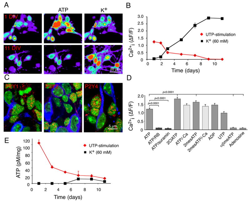
(A) Ca2+ responses of primary cortical neurons at 1 and 11 days in vitro (DIV) to ATP (100 μM) and K+ (60 mM). The cultures were loaded with the calcium indicator fluo-4/am (45 min, 4.6 μM). (B) Ca2+ responses to ATP and K+ as a function of DIV. means ± SEM (C) Expression of P2Y1, P2Y2, and P2Y4 (red) at 1 DIV. Expression of P2 receptors were lower in MAP2-positive neurons (blue) than in MAP2-negative cells. (D) Ca2+ responses to ATP and ATP analogs. The potency by which the ATP agonists mobilized intracellular Ca2+ stores is compatible with expression of functional P2Y1 (ADP), P2Y2 and P2Y4 (UTP) receptors. (E) ATP release in response to stimulation by UTP (100 μM) and high K+ (60 mM) as a function of DIV. The pseudocolor scale in A is similar to that in fig. 2B. Neurons were prepared from E16 mice pups, whereas neurospheres were prepared from E13 mice pups.