
Figure 3. Purinergic signaling sustained the proliferation of neural progenitors.
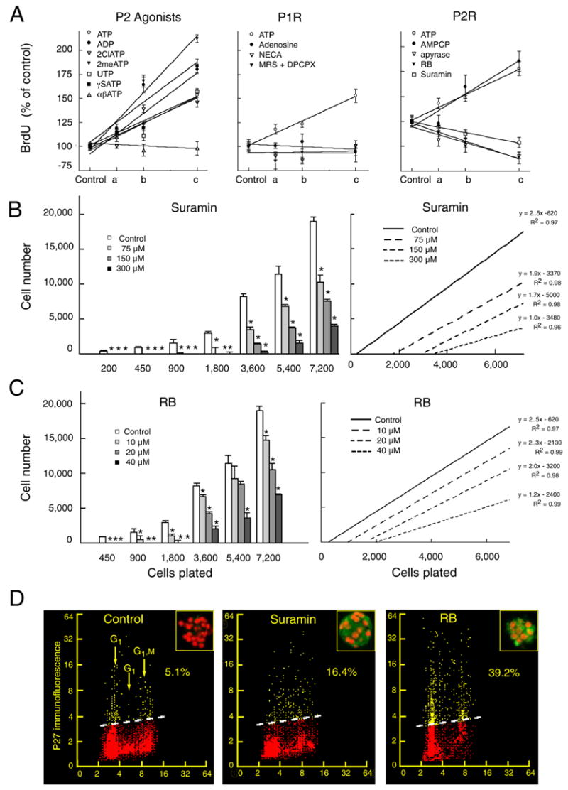
(A) The mitotic index of neural progenitors determined by the BrdU incorporation assay. The effect of ATP, ADP, 2ClATP, 2meSATP, UTP, γSATP, or αβATP upon BrdU incorporation relative to vehicle-treated controls was quantified at; 0 μM (control), 25 μM (a), 50 μM (b), and 100 μM (c) (left panel). Effect of ATP, adenosine, and NECA at; 0 μM (control), 25 μM (a), 50 μM (b), and 100 μM (c). A combination of the adenosine receptor antagonists DPCPX and MRS-1191 were tested at increasing concentration; 0 μM + 0 μM (control), 100 μM + 5 μM (a), 200 μM + 10 μM, and 400 μM + 20 μM (middle panel, same y-axis as left panel). Effect on BrdU incorporation of ATP or the ectonuclease inhibitor AMPCP; at 0 μM (control), 25 μM (a), 50 μM (b), and 100 μM (c). Apyrase was tested at: 0 U/ml (control), 10 U/ml (a), 20 U/ml (b), 40 U/ml (c). RB at; 0 μM (control), 10 μM (a), 20 μM (b), 40 μM (c). Suramin at; 0 μM (control), 75 μM (a), 150 μM (b), 300 μM (c) Lines represents 1-order linear regression. Regression coefficients were in the range of 0.89 to 0.99 (right panel). (B–C) Suramin and RB increased the minimal plating density required for survival of neural progenitors. At low plating densities, cells cultured in presence of suramin or RB died by 5 days in vitro *P < 0.01, one-way ANOVA, Bonferroni posthoc test. Right panels display 1-order regression analysis of the data in the left panel. (D) FAC analysis demonstrates an upregulation of the mitotic repressor, P27 in neural progenitor cells exposed to suramin (300 μM) and RB (40 μM). Bivariate distributions (scattergrams) representing DNA content (cell cycle distribution) versus expression of p27 in individual progenitor cells. The percent of p27 positive cells (above the threshold lines) was quantified based on the level of fluorescence of control cells stained with the secondary antibody only (isotypic control). Inserts Neural spheres immunostained against p27 (green). Nuclei are labeled with propidium iodide (red). Neurospheres were prepared from E13 mice pups.