


Fig. 2. Ability of various pancreatic secretagogues and growth factors to stimulate PKD1 S916 and S744/748 phosphorylation in rat pancreatic acini.
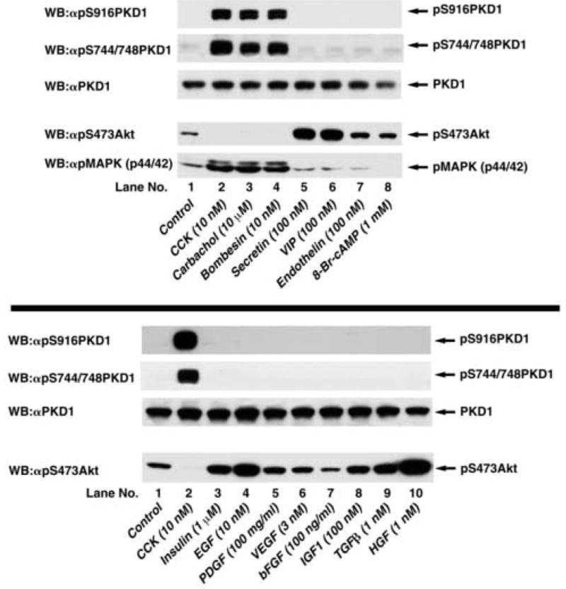
Top: Rat pancreatic acini were treated with no additions or with 10 nM CCK, 10 μM carbachol, 10 nM bombesin, 100 nM secretin or 100 nM VIP for 5 min, with 100 nM endothelin for 10 min, or with 1 mM 8-Br-cAMP for 30 min. Upper 3 panels: membranes were analyzed using either anti-pS916 PKD1 Ab (panel 1) or anti-pS744/748 PKD1 Ab (panel 2). To verify loading of equal amounts of protein, membranes were stripped and re-blotted with anti-PKD1 Ab (panel 3). Lower 2 panels: For positive controls for stimulation by pancreatic secretagogues, membranes were analyzed using anti-phospho Akt and anti-phospho MAPK Ab (panels 4, 5). The bands were visualized using chemiluminescence. Results from a representative experiment of 6 experiments are shown. Bottom: Rat pancreatic acini were treated with no additions, with 10 nM CCK for 5 min as positive control or with 10 nM EGF for 2.5 min, with 1 μM insulin or 100 mg/ml PDGF for 5 min, with 100 ng/ml bFGF or 1 nM HGF for 10 min, with 3 nM VEGF or 100 nM IGF1 for 15 min or with 1 nM TGFβ for 60 min. Upper 3 panels: membranes were analyzed using either anti-pS916 PKD1 Ab (panel 1) or anti-pS744/748 PKD Ab (panel 2). To verify loading of equal amounts of protein, membranes were stripped and re-blotted with anti-PKD1 Ab (panel 3). Lower panel: For positive controls for stimulation by growth factors, membranes were analyzed using anti-phospho Akt Ab (panel 4). The bands were visualized using chemiluminescence. Results from a representative experiment of 4 experiments are shown.