Fig. 1.
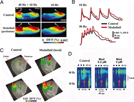
Modafinil enhances thalamocortical activity and reduces the edge effect in vitro. (A) Three-dimensional snapshots illustrate voltage-dependent fluorescence image spread, indicating increased cortical activity with paired white matter electrical stimulation at 40 Hz and 10 Hz (Left) or with only 10-Hz stimulation (Right) before (Upper) and after (Lower) 100 μM modafinil perfusion. (B) Pixel profiles recorded during 10-Hz (Upper) and 40-Hz stimulation (Lower) before (black) and after modafinil (red) (same slice as in A). (C) Three-dimensional VSDI elicited by thalamic VB 40-Hz paired shock stimulation before (control; Left) and after local micropressure (dotted lines; Right) of 100 μM modafinil. The VSDI results are superimposed on a phase-contrast image of the thalamocortical slice. (D) Profiles along cortical layer 5 after simultaneous 40-Hz (three pulses) and 10-Hz (two pulses) stimulation in control (Left) and 0.2 μM and 2 μM modafinil (Center and Right). Note the increased response resulting from the interaction of low and high frequencies after the second and third 40-Hz stimuli in the control panel (red arrow). Note how modafinil did reduce such activity (red arrows) (i.e., edge effect) (48).

