
Figure 3.
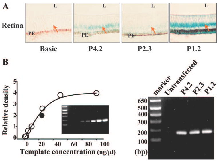
Expression of P4.2 reporter and deletion mutants in chicken retina. (A) X-gal staining of the retina of chicken embryo. P4.2 β-gal–Pax6 reporter and deletion mutants (P2.3 and P1.2) were introduced into the blastoderm of the chicken at E1. At E7, embryos were collected and stained with X-gal. Tissue sections were processed at a thickness of 10 μm. L and PE represent lens and pigment epithelial layer, respectively. (B) Copy numbers of the P4.2 reporter and mutants in transfected chicken embryos. Semi-quantitative PCR was performed with embryo DNA templates and a pair of primers specific to β-galactosidase gene sequences (180 bp apart). Chicken embryonic DNAs were isolated from the same individual embryos as shown in (A). The amount of PCR products as a function of template concentrations was plotted to generate a standard curve. DNA templates were diluted into 1, 2.5, 5, 10, 25, 50, and 100 ng/μL. There were at least six chicken embryos for each transfection experiment. Results from these experiments were very consistent.