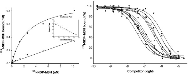
Figure 4.

Saturation curves with Scatchard plot and competition curves for the lamprey MCa receptors expressed in HEK-293 EBNA cells. The saturation curves (left) were obtained with 125I-labelled NDP-MSH and the figure shows total binding (filled square) and binding in the presence of 2 μM cold NDP-MSH (filled triangle). The lines represent the computer-modelled best fit of the data assuming that ligands bound to one site. The competition curves (right) for NDP-MSH (filled triangle, pointing up), α-MSH (filled square), β-MSH (open circle), γ1-MSH (open diamond), ACTH(1–17) (open square), ACTH(1–24) (filled circle), ACTH(1–39) (x), MTII (filled diamond), HS024 (open triangle, pointing down), MSH-A (filled triangle, pointing down), MSH-B (open triangle, pointing up) and ACTH (1–31) (asterisk) were obtained by using a fixed concentration of approx. 0.6 nM 125I-labelled NDP-MSH and varying concentrations of the non-labelled competing peptide.
