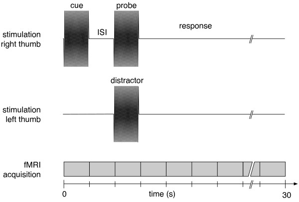
Figure 5.

Illustration of the experiment. Upper graph: A vibrotactile stimulus (frequency: 25 Hz; duration: 2 s) was delivered to the right thumb (cue) followed by an analogous probe of either identical frequency or higher frequency (25 Hz + individual discrimination threshold f). The interstimulus interval (ISI) was either 2 s (as illustrated here) or 8 s. Lower graph: In 25% of trials the probe was paired with a distractor to the left thumb. The stimulation parameters of the distractor were identical to those of the cue. Functional MRI data were obtained continously. Every 2 s, a brain volume consisting of 26 axial was acquired, starting with the beginning of each trial.
