
Figure 11.
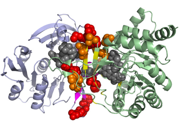
The important contacting residues across the PyrDB and the PyrK subunits of dihydroorotate dehydrogenase B (PDB code: 1EP1) assigned by our predictor. The high scoring residues at the binding site (yellow) of the PyrDB subunit (green) were colored orange and presented as spheres. The high scoring residues at the binding site (purple) of the PyrK subunit (light blue) were colored red and presented as spheres. Three cofactors, FMN, FAD and the [2Fe-2S] cluster were colored gray and presented as spheres.