
Figure 15.
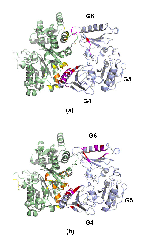
The important contacting residues across gelsolin G4–G6 domains and actin (PDB code: 1H1V) assigned by our predictor based on (a) known binding sites and (b) putative binding sites. The high scoring residues at the binding site (yellow) of actin (green) were colored orange. The high scoring residues at the binding site (purple) of gelsolin G4–G6 domains (light blue) were colored red.