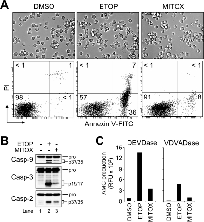
Figure 1. Topo2 inhibitors induce procaspase activation and apoptosis in wild-type Jurkat cells.
(A) Wild-type Jurkat cells (106/ml) were cultured with DMSO, 10 μM ETOP or 100 nM MITOX for 6 h and processed for light microscopy (40× objective) or the quantification of cell death by flow cytometric analysis of annexin V–FITC and PI staining as described in the Experimental section. Quadrants are defined as: live (lower left), early apoptotic (lower right), late apoptotic (upper right) and necrotic (upper left). Numbers refer to the percentage of cells in each quadrant. (B) and (C) Duplicate aliquots of cells were harvested and lysed for Western blotting or caspase activity measurements as described in the Experimental section. Enzyme activity was monitored by the production of AMC.

