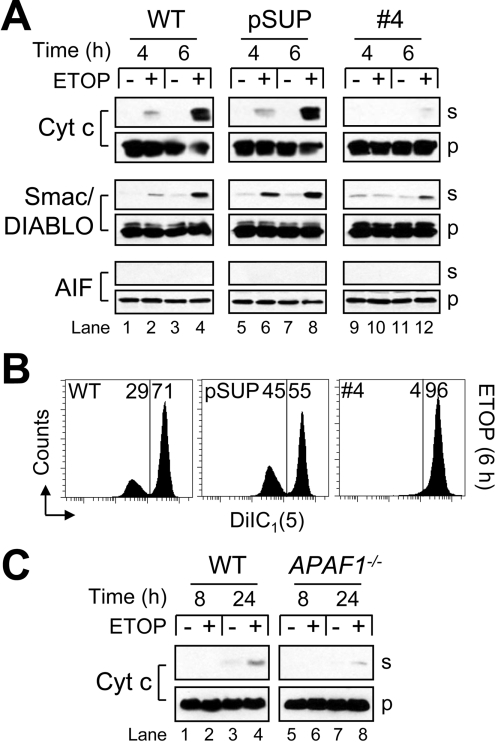
Figure 5. Impairment of mitochondrial apoptotic events in Apaf-1deficient cells.
(A) Wild-type (WT), control-vector-transfected (pSUP) and Apaf-1-deficient cells (106/ml) were cultured in the presence or absence of 10 μM ETOP for 4 or 6 h and harvested for subcellular fractionation as described in the Experimental section. Supernatant (s) and pellet (p) cell fractions were analysed by Western blotting. (B) Duplicate aliquots of cells at 6 h were processed for ΔΨ determination as described in the Experimental section. Reduced DiIC1(5) fluorescence is indicative of a loss of ΔΨ, and numbers refer to the percentage of cells in each region. (C) WT and APAF1−/− MEFs were cultured in the presence or absence of 50 μM ETOP for 8 or 24 h and harvested for subcellular fractionation. Supernatant (s) and pellet (p) cell fractions were analysed by Western blotting.

