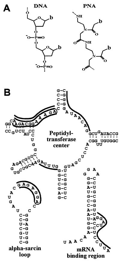
Figure 1.
(A) Chemical structure of a PNA oligomer compared with that of DNA. b indicates the nucleobases adenine, cytosine, guanine, thymine, or pseudo-isocytosine(20). (B) Target sites for antiribosomal PNAs. The binding sites for PNAs are shown as dark lines adjacent to the sequence. Triplex forming bis-PNAs (18) are shown connected by an ethylene glycol linker (thin line) (see also Table 1).

