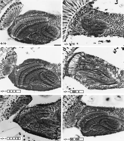
Figure 3.
Rescue of dock axon fiber pattern defects in the inner optic ganglia by mutant transgenes. (A) Horizontal section of a wild-type adult head stained with silver reveals the four highly ordered neuropil regions in the optic lobe, including the lamina (la), medulla (ome+ime), lobula (lo), and lobula plate (lp). R cell axon innervation induces the development of the outer optic lobe [i.e., lamina and outer medulla (ome)], but not the inner optic lobe [i.e., inner medulla (ime), lobula, and lobula plate] (51, 52). (B) In dockP1 mutants (−/−), neuropil structures were completely disrupted. Wild-type dock (C) or SH3–1/SH3–3 doubly mutant (F) transgenes rescued the optic lobe defects. (D) SH3–2 mutant transgene exhibits no rescue activity. (E) The SH2 mutant largely rescued the outer optic lobe, but not the inner optic lobe, defects. Genotypes: (A) Wild type; (B) dockP1. (C–F) Adult heads were from dockP1/dockP1 flies carrying one copy of elav-GAL4 transgene and the following UAS-transgene rescue constructs: (C) wild-type dock; (D) SH3–2 (W151K); (E) SH2 (R336Q); and (F) SH3–1 (W48K)/SH3–3 (W225K). Icons depict domain structure of Dock encoded by transgenes. (Left to right) Domains are SH3–1, SH3–2, SH3–3, and SH2. “X” indicates domain containing mutation. (Bar = 20 μm.)

