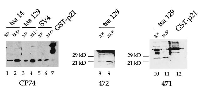
Figure 1.
Expression of p21 by Western blot analysis. Total protein (30 μg) extracted from tsa14, 129, and SV4 cell lines cultured at 33°C and at 39.5°C were analyzed by immunoblotting using CP74, a mAb specific for p21 (lanes 1–7; provided by E. Harlow, Massachusetts General Hospital). Two affinity-purified rabbit polyclonal peptide antibodies, 471 and 472 (purchased from Santa Cruz Biotechnology), were also used to analyze extracts prepared from tsa129 cells (lanes 8–12). Enhanced chemiluminescence system was used for detection. The GST–rat p21 fusion protein was used as positive control. Surprisingly this fusion protein is recognized by the 471 antibody whereas endogenous p21 is not detected.

