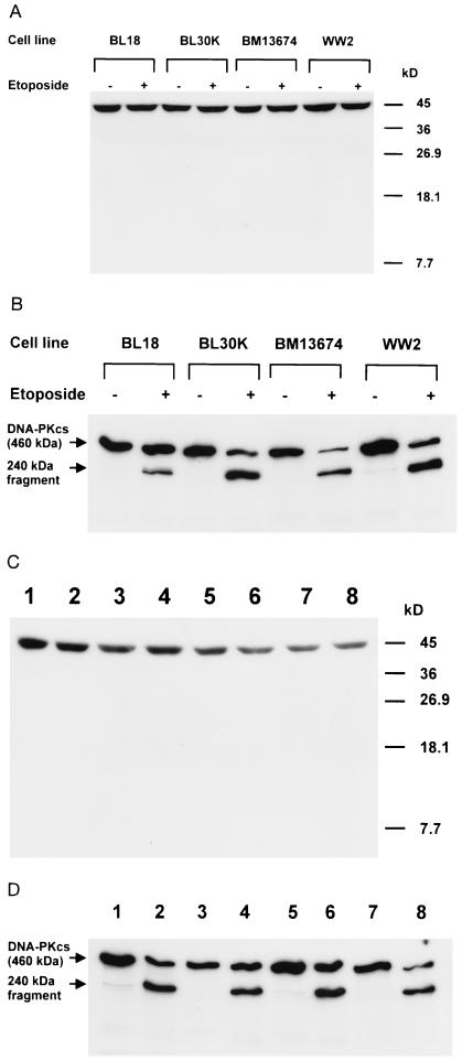
Figure 4.
Lack of actin cleavage in different cell types by a variety of agents causing apoptosis. (A) Lack of cleavage of actin in different Burkitt lymphoma cells after exposure of cells to 40 μM etoposide. A polyclonal antibody against the C-terminal 11 amino acids of actin was used for immunoblotting. (B) Cleavage of DNA-PKcs during apoptosis induced by etoposide (40 μM) in different Burkitt lymphoma cells. DNA-PKcs was detected with DPK1 antibody. (C) Lack of degradation of actin in HeLa, Molt-4, U937, and CTL cells undergoing apoptosis induced by different agents. For HeLa cells, extracts were prepared from detached cells after 8 h of treatment with etoposide (40 μM). For Molt-4 cells, cells were irradiated (20 Gy) and extracts were prepared 8 h after irradiation. For U937 cells, cells were pretreated with interferon γ (200 international units/ml) for 24 h and then incubated with anti-Fas antibody at 50 ng/ml for 10 h prior to preparation of extracts. CTLs were incubated with specific peptide for 4 h prior to preparation of extracts. Lane 1, Molt4 untreated; lane 2, Molt4 exposed to 20 Gy of γ-irradiation; lane 3, HeLa untreated; lane 4, HeLa + 40 μM etoposide; lane 5, CTL untreated; lane 6, CTL + specific peptide; lane 7, U937 untreated; lane 8, U937 + 50 ng/ml anti-Fas antibody. A polyclonal antibody against the C-terminal 11 amino acids of actin was used for immunoblotting. (D) Cleavage of DNA-PKcs during apoptosis induced by different agents in HeLa, Molt-4, U937, and CTL cells. DNA-PKcs was detected with DPK1 antibody.

