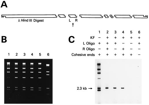
Figure 2.
Sequence-specific labeling of a fragment of λ DNA. (A) Schematic showing the position of the 2.3-kb λ DNA HindIII fragment labeled. The arrow points to the 2.3-kb fragment, and L and R show the positions of the two oligonucleotides used to direct sequence-specific ligation to a short radioactive duplex with a HindIII cohesive end. (B) Agarose gel stained with ethidium bromide showing the HindIII fragments of λ DNA in lanes 1–5 and a ScaI digest in lane 6. The two shortest HindIII fragments have run off the bottom of the gel. (C) Autoradiogram of the dried gel. Lane 1 shows λ DNA where KF was omitted and every fragment was labeled. The band at 4.4 kb is less intense because of ligation to the 23.1-kb band via the terminal λ cos sites. Lane 2 shows labeling of both ends of the 2.3-kb fragment. Lanes 3, 4, and 5 show the effect of omitting either one or both of the oligonucleotides. Lane 6 shows the results by using λ DNA fragments with blunt ends.

