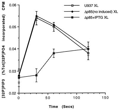
Figure 3.
Expression of a dominant-negative form of p85, Δp85, abolishes only the early peak of PIP3 observed after FcγRI aggregation. The time course of FcγRI-stimulated PIP3 concentrations were measured in cells induced to express to dominant-negative form of p85 (Δp85 + IPTG XL) and compared with the same transformed cells not induced to express [Δp85 (no induced) XL] and wild-type U937 cells (U937 XL). Data are the mean ± SD of triplicate measurements for each time point and are derived from three separate experiments.

