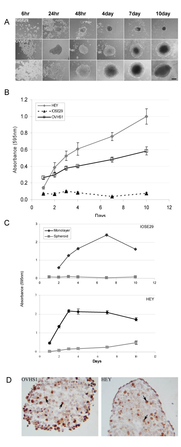
Figure 1.
(A) Formation of normal and ovarian carcinoma cell line spheroids over 10 days. HEY, OVHS1 and IOSE29 cell lines at a density of 104cells/ml were seeded on 0.5% agarose-coated wells in the presence of normal growth medium for 10 days. Aggregation of cells was viewed using an inverted microscope with phase contrast, magnification 100 ×. (B) Proliferation of ovarian carcinoma cells grown as spheroids. HEY, OVHS1 and IOSE29 cells were seeded on agarose-coated plates as described in the Materials and Methods. The level of proliferation was measured by MTT assay as described in the Materials and Methods. Data are representative of three experiments expressed as mean ± SEM of twelve replicates. (C) Comparison of proliferation of IOSE29 (upper panel) and HEY (lower panel) cells grown as monolayer versus spheroids. Proliferation was measured by MTT assay as described above and data are expressed as mean ± SEM of six replicates. (D) Immunohistochemical staining of Ki67 in day 4 HEY and OVHS1 spheroids. Day 4 HEY and OVHS1 spheroids were collected, embedded in OCT, sectioned and stained as described in the Materials and Methods. Magnification 400 ×.

