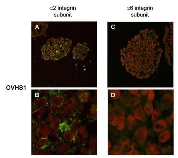
Figure 7.
Cellular localisation of α2 and α6 integrin subunits in spheroids grown for 4 days. Using Alexa-fluor immunofluorescence, spheroids embedded in OCT were sectioned and stained for α2 and α6 integrin subunits and counterstained as described in the Materials and Methods. Images captured at magnification 400 × using an oil immersion lens on a Leica con-focal microscope. B and D are 1.5 × magnification of A and C respectively

