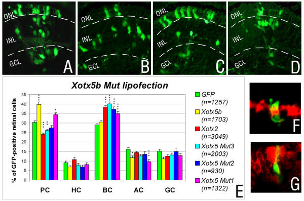
Figure 2.
Results of in vivo lipofection of RPCs with wild-type Xotx2 and Xotx5b, and mutant Xotx5b constructs. (a-d) Sample sections are shown for control retinae lipofected with GFP+vector DNA alone (a), GFP+Xotx2 (b), GFP+Xotx5b (c) or GFP+Xotx5bMut3 (d); GCL, ganglion cell layer; INL, inner nuclear layer; ONL, outer nuclear layer. (e) Overall distribution of retinal cell types in clones lipofected with the different constructs; PC, photoreceptor cells; HC, horizontal cells; BC, bipolar cells; AC, amacrine cells; GC, ganglion cells. The proportion of each cell type is represented as an average. Error bars indicate the standard error of the mean. The experiment was repeated at least three times for all constructs. Counted cells are indicated in the histogram (n), from 15 retinae for GFP, 15 retinae for Xotx5b, 18 retinae for Xotx2, 16 retinae for Xotx5bMut3, 10 retinae for Xotx5bMut2, and 13 retinae for Xotx5bMut1. Asterisks represent significant differences between Xotx constructs and GFP, as calculated by ANOVA analysis using the Tukey-Kramer post-test (*p < 0.05, **p < 0.01, ***p < 0.001). (f, g) In situ hybridization analyses showing examples of GFP-positive (green), Xotx5-lipofected photoreceptor cell positive for IRBP probe (Fast Red detection) (f), and a Xotx5bMut3-lipofected bipolar cell expressing Xvsx1 (Fast Red detection) (g).

