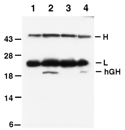
Figure 6.
Immunoblot analysis of anti-hGH immunoprecipitates from sera of hGH transgenic mice and from the spent medium of keratinocytes cultured from newborn animals. Sera were taken from the bloodstream of 9-month-old transgenic and control mice. Keratinocytes from newborn control and transgenic mice were cultured in duplicate as described; at 80% confluence, fresh medium added, and the medium was sampled after an additional 24 hr of culture. Immunoprecipitations to detect the hGH transgene product in the skin or in the culture medium were carried out using mAb-coated plastic beads for hGH (Hybritech) under the same conditions as RIAs (see Materials and Methods), but without radiolabeled tracer. Aliquots of the immunoprecipitates were then resolved by SDS/PAGE and subjected to immunoblot assays, performed with a mAb against hGH (Sigma). Immunoprecipitates were from control serum (lane 1), transgenic serum (lane 2), control medium (lane 3), and transgenic medium (lane 4). Migration of molecular mass standards are indicated at left in kilodaltons; migration of heavy (H) and light (L) chains of γ-immunoglobulin is given at right.

