
Figure 5.
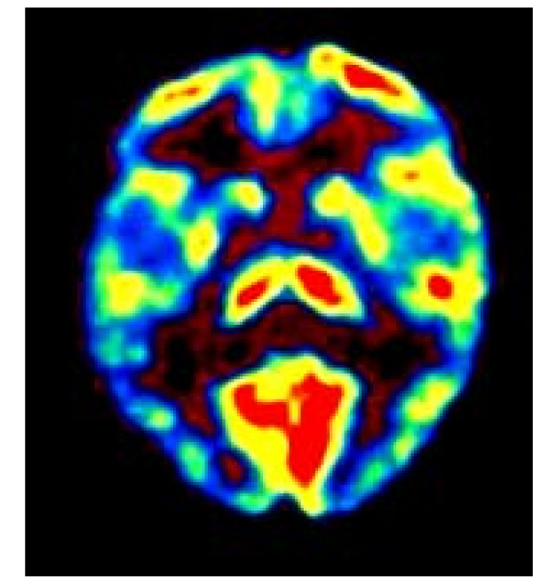
FDG-PET of patient #1. [18F] deoxyglucose (FDG) positron emission tomography (PET) had been performed in each patient before treatment. The area of hypermetabolic activity in the temporal cortex was chosen as target for TMS treatment. Here the FDG PET of patient #1 is displayed, where a transversal slice through the temporal brain region shows unilaterally increased metabolic activity in projection to the left auditory cortex.