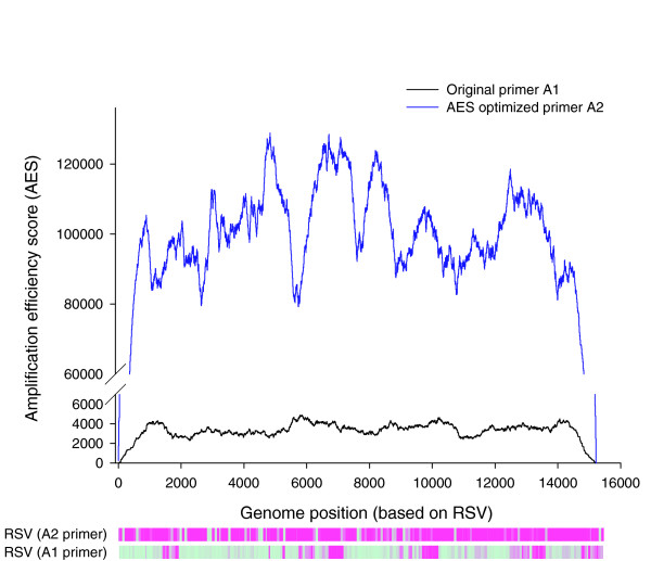
Figure 3.

Measurement and application of AES. An RSV patient sample was amplified using original primer A1 (black line), or AES-optimized primer (blue line). The probes that have detectable signal above threshold are shown in purple in the corresponding heatmaps. For primer A1, the detectable regions correspond to regions that have higher AES scores than undetectable regions.
