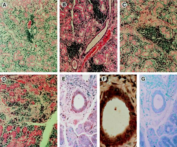
Figure 2.

Microscopic examination of the salivary glands from HCV envelope transgenic mice. (A) A 3-month-old male from the E101 line. (B) A 6-month-old male from the E139 line. (C) A 16-month-old male from the E101 line. (D) A 16-month-old female from the E139 line. (E) Immunostaining of the E1 protein; a 9-month-old male from the E101 line. (F) A higher magnification of E. (G) A control staining of E. Paraffin-embedded sections of salivary glands were stained by hematoxylin and eosin (A–D) or immunostained with anti-E1 rabbit serum (E and F) or preimmune rabbit serum (G) by the avidin–biotin complex method (28) and counterstained by methyl green (E–G). (A–D, ×80; E and G, ×200; F, ×500.)
