Fig. 3.

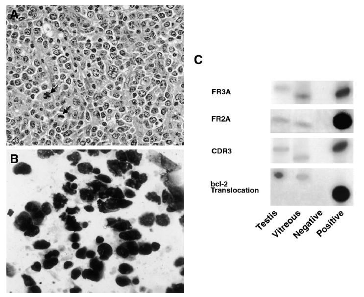
Case 1. A: Histopathology of the testicular biopsy specimen showing atypical lymphoid cells with multiple mitotic figures (arrows) (hematoxylin & eosin, original magnification, ×400). B: Cytology of the vitreous specimen showing atypical lymphoid cells with enlarged hyperchromatic nuclei and scant cytoplasm (giemsa, original magnification, ×640). C: Polymerase chain reaction amplification of microdissected cells from the testicular and vitreous specimens showing IgH gene rearrangements of the CDR3 region with three different primer sets. There are different patterns of IgH gene rearrangements at the CDR3 sites between the testicular and intraocular lymphomas. The testicular and vitreous samples were also positive for the bcl-2 t(14;18) translocation.
