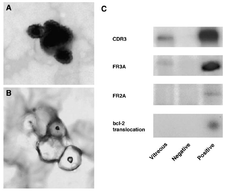
Fig. 4.

Case 2. A: Histopathology of the vitreous specimen showing large lymphoid cells with scanty cytoplasm, prior to microdissection (giemsa, original magnification, ×400). B: Histopathology of the same vitreous specimen post microdissection (giemsa, original magnification, ×400). C: Polymerase chain reaction amplification of microdissected cells from the vitreous specimen showing a positive IgH gene rearrangement at the CDR3 site with the FR3A primers. The vitreous sample was negative for the bcl-2 t(14;18) translocation.
