
Figure 4.
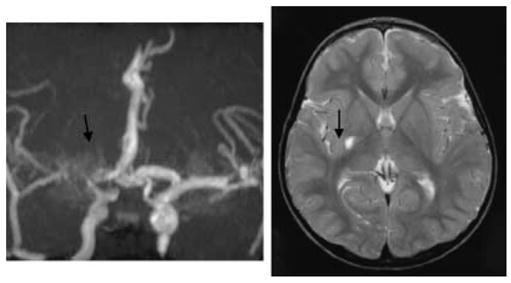
Magnetic resonance angiogram (MRA) and imaging (MRI) from a girl (Patient 10, Tables 2 and 3) with sickle cell anaemia who had a sleep study showing dips and high transcranial velocities (up to 267 cm/second) for 8 years without symptoms but presented with a hemiparesis in the context of acute aplastic anaemia secondary to Parvovirus. Although the MRA shows severe turbulence with some ‘moyamoya’ collaterals (arrow), the MRI shows only a small infarct (arrow) and the patient made a complete recovery. This might be an example of the preconditioning effect of prior exposure to hypoxia in limiting the extent of tissue injury at the time of acute stroke but at the cost of irreversible vascular change.