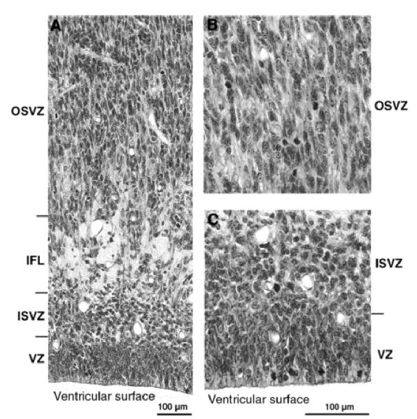
Figure 5.

Photomicrographs at higher-power of the germinal zones at E78. (A) Note the radial texture of the dominant OSVZ and of the lesser VZ. Note also the IFL separating the radial OSVZ from the more irregularly orientated nuclei of the ISVZ. (B) Higher-power view of the OSVZ. Note radial texture of tissue and the presence of mitotic figures. (C) View of the ISVZ and VZ. Note the presence of mitotic figures among the irregularly orientated nuclei of the ISVZ. In the VZ, nuclei are radially arranged and mitotic figures lie towards the ventricular surface, indicating the continuation of interkinetic nuclear migration.
