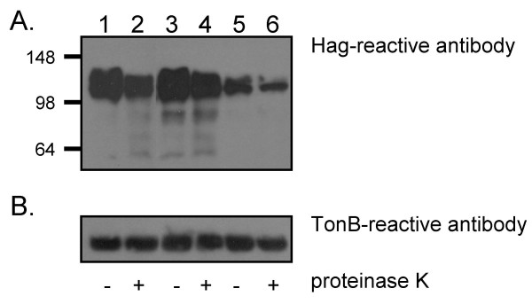
Figure 5.

Western blot analysis of recombinant E. coli cells treated with proteinase K. E. coli carrying the plasmids pELO35.Hag (lanes 1 and 2), pBBHS2.24 (lanes 3 and 4) and pBBHS3.20 (lanes 5 and 6) were incubated for 15 min on ice in the presence (lanes 2, 4 and 6) or absence (lanes 1, 3 and 5) of proteinase K. These cells were lysed, resolved by SDS-PAGE, transferred to PVDF membranes and probed with the anti-Hag antibody 5D2 (panel A) or anti-TonB antibody 4F1 (panel B). Molecular weight markers are shown to the left in kDa.
