
Figure 3.
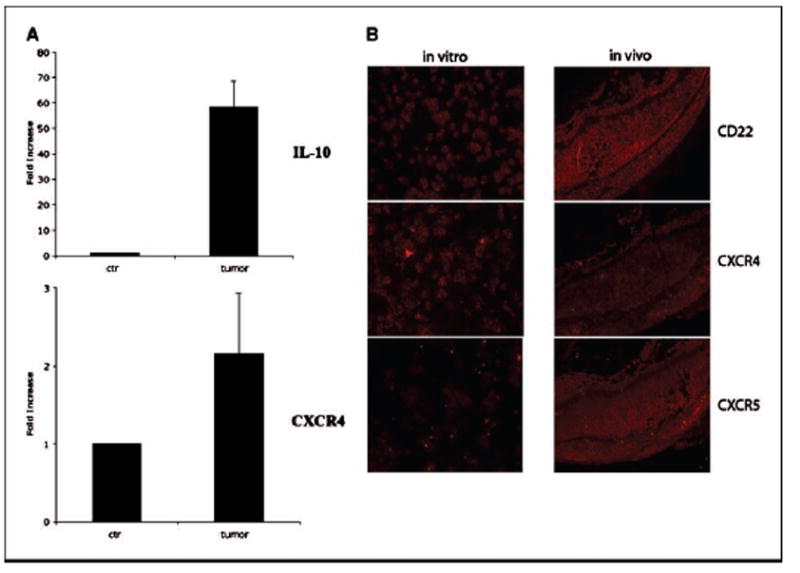
Molecular mimicry of mouse intraocular B-cell lymphoma model to human PIOL. CA46 cells were intravitreally injected into SCID mice (20,000 per eye per injection), animals were sacrificed on day 21 postinjection, and eyes were collected and snap-frozen on dry ice. The frozen eye tissues were sectioned and stained for tumor cells. The tumor cells were microdissected out from the eye section, total RNA was isolated, and real-time PCR was done as described in Materials and Methods. A, in vivo expression of both IL-10 (top) and CXCR4 (bottom) in the tumor cells. B, immunohistochemistry shows in vivo expression of CD22, CXCR4, and CXCR5 on the tumor cells. Left, expression levels of CD22, CXCR4, and CXCR5 in the in vitro cultured tumor cells. Right, in vivo expression levels in the mouse tissue after injection on day 21 (red, positive staining). All molecular markers are expressed in vivo and the expression of CXCR5 seems to be up-regulated in vivo (see Results and Discussion for details). Overlay pictures showing both DAPI staining (blue, nuclei) and the antibody staining (red, CD22, or CXCR4 or CXCR5) can be seen in Supplementary Fig. S1.