
Figure 1.
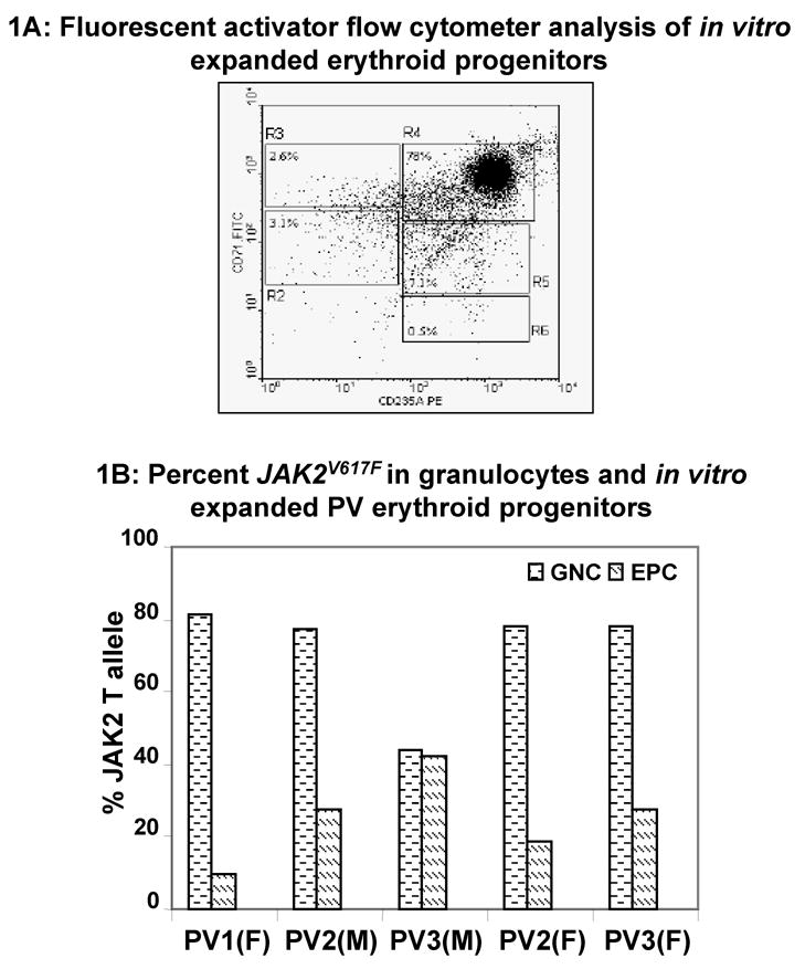
(A) Fluorescent activator flow cytometer analysis of in vitro expanded erythroid progenitors: Mononuclear cells were isolated from PV patients and expanded as described in the Methods. About 5 × 10⁵ cells were stained with PE-conjugated anti-CD235A antibodies (glycophorin) and FITC-conjugated anti-human-CD71 antibodies (transferrin receptor). Regions R2 to R6 are defined by characteristic staining pattern of erythroid cells. These are primitive progenitor cells plus mature BFU-Es and CFU-Es in R2 (CD71med CD235Alow), pro-erythroblasts and early basophilic erythroblasts in R3 (CD71high CD235Alow), early and late basophilic erythroblasts in R4 (CD71high CD235Ahigh), polychromatophilic and orthochromatophilic erythroblasts in R5 (CD71med CD235Ahigh), and late orthochromatophilic erythroblasts and reticulocytes in R6 (CD71low CD235Ahigh). (B) Percent JAK2V617F in granulocytes and in vitro expanded erythroid progenitors: Granulocytes (GNC) were isolated from peripheral blood of PV patients. JAK2V617F was measured in genomic DNA of granulocytes as well as in vitro expanded erythroid progenitors. M and F represent male and female patients, respectively.