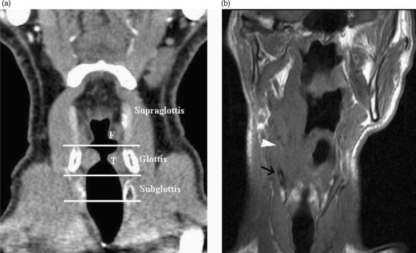
Figure 2.
(a) Coronally reformatted CT demonstrates the supraglottis, glottis and subglottis as defined by the level of the ventricular complex (false cords (F), true cords (T) and intervening ventricle). (b) T1-w coronal image demonstrates a supraglottic tumour extending inferiorly to ventricular level (arrowhead). Low T1-w signal (arrow) in the thyroid cartilage is non specific for reactive change or tumour invasion.

