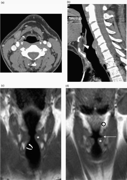
Figure 3.
(a) Axial, (b) sagittal reformatted CT images with (c) and (d) T1-w coronal MR images illustrating the submucosal laryngeal spaces. (a) The pre-epiglottic space (open arrowhead) and paraglottic space (white arrowhead) are seen in continuity. (b) More superiorly the pre-epiglottic space (white arrowhead) is bound anteriorly by the thyrohyoid membrane (see also (a)) whereas the thyroid cartilage is present anteriorly at a more inferior level (see also Fig. 1a). The epiglottis is present posteriorly. Soft tissue seen within the superior aspect of the pre-epiglottic space (open arrowhead) corresponds to the hyoepiglottic ligament. (c) The medial relations of the paraglottic fat (o) are seen to be the vestibule, the ventricle (long arrow) and the conus elasticus more inferiorly (curved arrow). Laterally it is related to thyroid cartilage and cricothyroid membrane. The true vocal cord is indicated (*).

