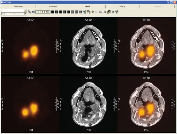
Figure 4.
Example of alternating fused images visualised in a PACS (AGFA Web 1000). Transverse slices of the neck region are shown in fused SPECT (indium-111 octreotide) and MRI (t1-weighted sequence) images. The series consists of three alternating representations of each slice: separate SPECT (left), separate MRI (middle) and combined SPECT-MRI (right). The MRI is difficult to interpret due to multiple surgery and the presence of metal. The SPECT shows suspected regions for remaining tumour tissue. The fused images provide the location of these regions.

