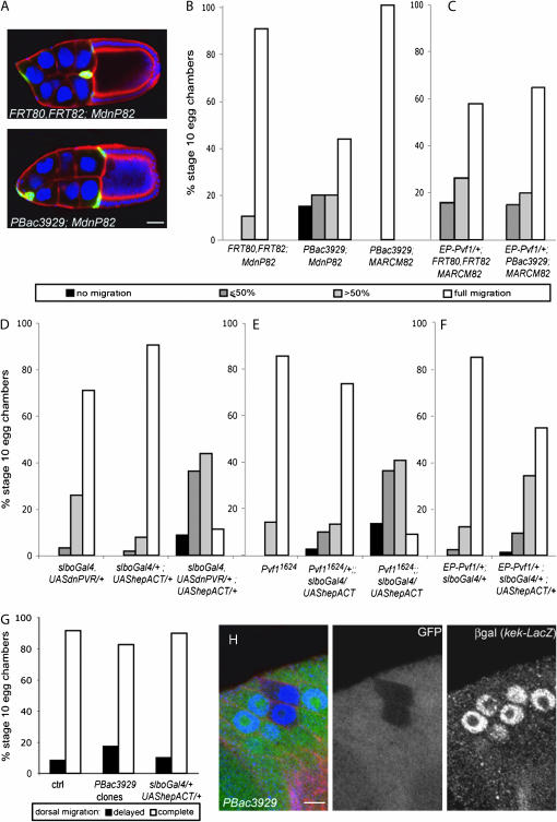
Figure 3.—
puc is required in border cells with low PVR signaling. (A) Stage 10 egg chambers, stained with DAPI (blue) to show nuclei, anti-β-galactosidase antibody (green) to reveal mutant cells, and phalloidin (red) from females of the following genotypes: hsFLP,UAS-lacZ/+; slbo-Gal4,UAS-dnPVR/+; FRT80,FRT82/FRT82,tub-Gal80 (top) and hsFLP,UAS-lacZ/+; slbo-Gal4,UAS-dnPVR/+; FRT80,FRT82, pucPBac3929/FRT82,tubGal80 (bottom). Bar, 20 μm. (B–F) Quantification of border cell migration toward the oocyte (posterior migration) in stage 10 egg chambers. (B) pucPBac3929 clones with or without dnPVR and control MdnP clones from females of the genotypes hsFLP,UAS-lacZ/+; slbo-Gal4,UAS-dnPVR/+; FRT80,FRT82/FRT82,tub-Gal80 (left, n = 10); hsFLP,UAS-lacZ/+; slbo-Gal4,UAS-dnPVR/+; FRT80,FRT82, pucPBac3929/FRT82,tub-Gal80 (middle, n = 21); and hsFLP,UAS-GFP/+; tub-Gal4/+; FRT80,FRT82, pucPBac3929/FRT82,tub-Gal80 (right, n = 10). (C) Control border cells overexpressing Pvf1 (left; genotype hsFLP,UAS-GFP/EP-Pvf1; tubGal4/+; FRT80,FRT82/FRT82,tubGal80, n = 19) and pucPBac3929 clones overexpressing Pvf1 (right; genotype hsFLP,UAS-GFP/EP-Pvf1; tubGal4/+; FRT80,FRT82,pucPBac3929/FRT82,tubGal80; n = 20). (D) Border cell clusters expressing dnPVR, hepACT, or both (n = 93, 272, 80). (E) Clusters expressing hepACT in control, Pvf1/+, or Pvf1 mutant background (n = 42, 149, 22). (F) Clusters expressing uniform Pvf1, alone or with hepACT (n = 203, 73). Genotypes are indicated and include the controls in each experiment. (G) Quantification of dorsal migration in pucPBac3929 border cell clones (from hsFLP/+; FRT80,FRT82, pucPBac3929/FRT82,UbiGFP females, n = 23), border cells expressing hepACT (slbo-Gal4/+; UAS- hepACT/+, n = 20), and the control (slbo-gal4/+, n = 12). (H) Follicular epithelium of stage 10 egg chamber from female of the genotype hsFLP/+; kek-lacZ/+; FRT80,FRT82, pucPBac3929/FRT82,UbiGFP, stained with anti-β-galactosidase antibody (β-gal, blue in overlay) to reveal kek-lacZ reporter expression and phalloidin (red in overlay). pucPBac3929mutant cells are marked by absence of GFP (green in the overlay). Dorsal is to the top. Bar, 5 μm.

