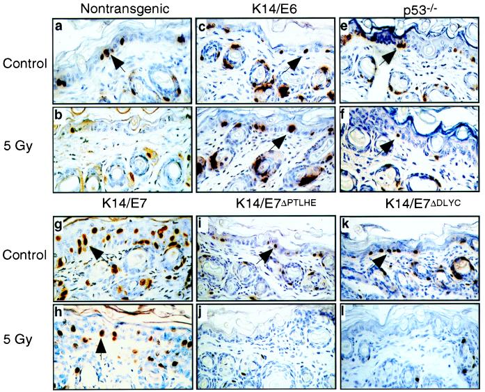
Figure 1.
Comparison of levels of DNA synthesis in nontransgenic, E6-transgenic, p53-null, and E7 transgenic epidermis after treatment with radiation. Shown are high-power magnification (×400) images of cross sections of skin from nontransgenic, K14HPV16E6 transgenic, p53−/−, K14HPV16E7, K14HPVE7ΔPTHLE, and K14HPV16E7ΔDLYC transgenic mice stained immunohistochemically for BrdUrd. Mice were either not treated (a, c, e, g, i, and k) or treated with 5 Gy of ionizing radiation 24 hr prior to sacrifice (b, d, f, h, j, and l). Arrows indicate examples of BrdUrd-positive (brown-stained nuclei) cells in the epidermis.

